


Research
Regulatory Mechanism of the Vertebrate Development
Throughout the vertebrate development, individual embryonic cells undergo a variety of processes (cell division, migration, differentiation, apoptosis etc.), and are finally strictly organized into a living organism It is now considered that this process is driven by ordered expression of a great number of genes within the cell nucleus Meanwhile, it is established that a variety of interaction between cells or tissues play important roles within individual embryos for establishment of the body plan at early stages and formation of many organs (organogenesis) and the entire organism at later stages.
Our current main research interest is on the molecular mechanism governing the regionalization of the vertebrate brain The vertebrate brain is one of the most complicated structures that can be seen in nature, and its development is an extraordinarily interesting issue, which is also really important from clinical reasons After neural induction in early embryos by the Spemann organizer, the induced neural region forms the neural plate, in which the anterior-posterior (AP) axis is established under the influence of posteriorizing signals emanating from the posterior region Succeedingly, a number of signaling centers are newly generated within the neural plate, such as the midbrain-hindbrain boundary (MHB), anterior neural ridge (anterior boundary cells), and the 4th rhombomere in the hindbrain Local signal centers thus formed pattern the adjacent brain primordia, leading to further regionalization of the embryonic brain In parallel, the dorsoventral (DV) polarity of the neural plate/tube is provided by the reciprocal signals from the notochord and epidermal region These complicated patterning processes along the AP and DV axes provide the framework for the 3-dimensional structure of the embryonic brain which finally develops an highly organized vertebrate brain with magnificent higher-order functions.
We are seeking to reveal the molecular mechanism underlying the regionalization of the brain, especially placing a focus on the formation of the midbrain-hindbrain boundary (MHB) and its adjacent brain regions The MHB is one of the well studied local signal centers generated at the future border between the midbrain and hindbrain, which generate signals to pattern the midbrain (tectum and tegmentum) and anterior hindbrain (future cerebellum) Previous studies have shown that MHB is induced by interaction within the neural plate or between the neural plate and the underlying mesoderm Furthermore, a number of genes have been identified as involved in the establishment and following development of the MHB, including transcriptional regulatory genes such as otx2, gbx2, pax2, engrailed, as well as secretory-factor genes such asfgf8 and wnt1 So MHB development can be regarded as the typical developmental process driven both by tissue interaction and regulation of regulatory genes that are spatially and temporally strictly regulated Aiming at solving these issues, we are using the zebrafish, Danio rerio, as the experimental model of vertebrate development.
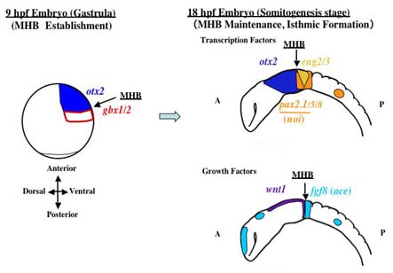
Fig. 1. Regulatory genes involved in the development of the midbrain-hindbrain boundary.
The main research projects are as follows:
1. Roles of the gbx homeobox genes in the development of the MHB region.
Roles of the gbx homeobox genes in the development of the MHB region
gbx1 and gbx2 encode Gbx-type homeodomain transcription factors that are conserved throughout the animal kingdom Our previous study (Kikuta et al., 2003) as well as others showed that gbx genes are involved in the development of MHB at early and late stages of development in all the vertebrates examined We are addressing the molecular aspects of the roles played by these two genes in the embryonic vertebrate brain.
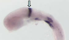
Fig. 2. Expression of gbx2 in zebrafish embryos at 24 hours post-fertilization The expression of gbx2 is marked with an arrow.
2. Roles of pou2 in the development of MHB and other embryonic regions
pou2 is a zebrafish POU-type homeobox gene that was found as regulating the development of MHB and the hindbrain Its mammalian homologue, Oct3/4, is known to be involved in the regulation of cell differentiation at early stages in mice as well as of germ cell development We are examining the molecular mechanism of the pou2 functions that are seen in many aspects of zebrafish development.
3. Transcriptional regulation of the genes regulating brain development.
Regulatory genes involved in regionalization of the brain are expressed specifically in most cases in terms of the embryonic region and stage To understand the molecular mechanism conferring positional values to the neural plate, therefore, we are examining the transcriptional regulation of several brain regionalizing genes expressed in the vicinity of MHB, such as gbx2,fgf8, hoxb1b, and pou2* In this respect, the zebrafish is an excellent animal that allows efficient establishment of transgenic lines and observation of the expression of fluorescent reporter genes Indeed, we have identified a number of regulatory regions in the vicinity of genes of interest, and found out several regulatory transcription factors for these brain-forming genes (Inoue et al., 2006; Islam et al., 2006).
 |
Fig. 3. Expression of green fluorescent protein (GFP) that is driven by the regulatory region of a certain brain forming gene. |
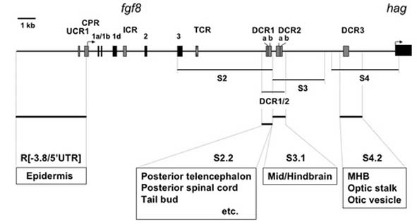 |
Fig. 4. Regulatory regions identified in the vicinity of zebrafishfgf8 (Inoue et al., 2006) |
*gbx2 andfgf8 are specifically expressed in the anterior-most hindbrain and regulate the MHB development. In addition,fgf8 is expressed in other signaling centers, contributing to the pattering of a number of embryonic regions and organs. In most cases fgf8 is considered one of the most dominant regulatory growth factors in patterning and organogenesis. pou2 is expressed transiently around the end of gastrulation broadly in the brain from the midbrain to the hindbrain, patterning these brain regions. Meanwhile, hoxb1b is a functional homologue of mammalian Hoxa1, and is expressed in the posterior neural region up to the r3/r4 boundary, providing the positional cue for establishing r4, which is the signal center regulating the hindbrain.
4. Roles of the FGF signaling in zebrafish development.
In mammals, 22 FGF (fibroblast growth factor) members have been identified and are known to be among the most important regulators in many aspects of vertebrate development. In early embryos, FGF signals are involved in induction of the mesoderm and neural region, as well as in the establishment of the dorsoventral axis of the body Later, it contributes to patterning of a number of organs including the brain, limb, bone, and visceral organs. To reveal their roles in early development and brain regionalization, especially in the prospective midbrain, MHB, and hindbrain, we are examining the functions of the four FGF receptors (Fgfr1-4) and modifying factors of the FGF signals in zebrafish development (Tonou-Fujimori et al, 2002).
5. Screening of novel regulatory genes of brain formation by mutagenesis.
We have conducted a mutagenesis screening of zebrafish mutants with defects in brain formation, and obtained several lines with interesting phenotypes Mapping of one of the mutations is now in progress.